The elbow is a junction between the forearm and the upper arm. The elbow joint is made up of 3 bones namely the humerus bone in the upper arm which joins with the radius and ulna bones in the forearm. The elbow joint is essential for the movement of your arms and to perform daily activities. The head of the radius bone is cup-shaped and corresponds to the spherical surface of the lower end of the humerus. The injury in the head of the radius causes impairment in the function of the elbow. Radial head fractures are very common and occur in almost 20% of acute elbow injuries. Elbow dislocations are generally associated with radial head fractures. Radial head fractures are more common in women than in men and occur more frequently in the age group of 30 and 40 years.
Causes
The most common cause of a radius head fracture is breaking a fall with an outstretched arm. Radial head fractures can also occur due to a direct impact on the elbow, a twisting injury, sprain, dislocation or strain.
Symptoms
The symptoms of a radial head fracture include severe pain, bruising and swelling in the elbow, and difficulty in moving the arm. Dr. Bala will ensure that you do not have any symptoms in the forearm or wrist.
Diagnosis
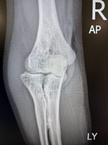
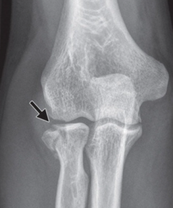
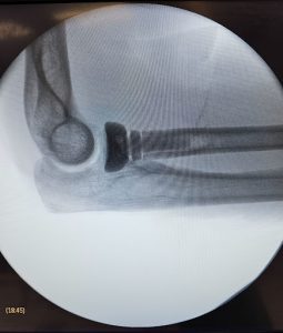
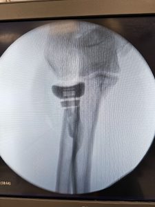
An X-ray is required to confirm the fracture and assess displacement of the bone. Sometimes, Dr Bala might suggest a CT scan may also be needed to obtain further details of the fracture, especially the joint surfaces. Dr Bala might inject local anaesthesia into the joint and check for a block to forearm rotation movements to decide on the need for surgery. He may also ask for x-rays of the entire forearm or of the wrist of the same side to rule out associated injuries.
Treatment
The treatment of a fracture depends on the type of fracture.
- Type 1 fractures are usually very small. The bone appears cracked, but remains together. It is common to wear a sling or cast for a few days. If the fracture becomes displaced then surgical intervention may be required.

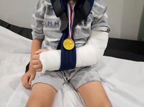
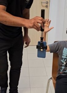
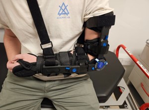
- The aim of treatment is to maximise early motion and to reduce the risk of stiffness. Non-surgical treatment options include pain medication, ice application, the use of a splint or a sling to immobilise the elbow during the healing process and physiotherapy.
- Type 2 fractures are characterised by fracture displacement treated with surgery. During surgery, headless titanium screws or very thin plates may be inserted to hold the displaced bone together in an inverted tripod fashion. Small pieces of bone may be removed (radial head partial excision) if it prevents normal movement of the forearm. Dr Bala prefers very low-profile plates, if at all, in this region as they may be bulky, restrict normal forearm rotation and often need another surgery to be removed.


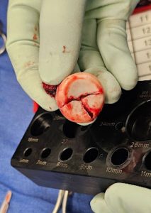
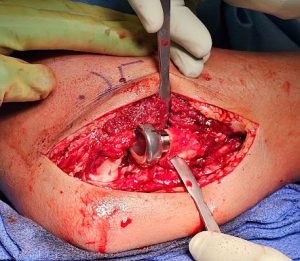
- Type 3 fractures are characterised by multiple fractures/pieces of the radial head. Surgery is recommended to either fix or replace the radial head with an artificial radial head replacement. When performing a radial head replacement Dr Bala prefers a prosthesis that is modular, made of cobalt chromium alloy and has a short, straight stem. Depending on the associated injuries Dr Bala may approach this fracture either from the outside of the elbow or from the back. He uses a ligament sparing approach to protect important ligaments on the outer side of the elbow.
- Radial head replacements if not properly sized or done without adequate ligament repair may predispose to stiffness, laxity or arthritis in the long term. They may sometimes need removal after many years if problems arise.


Dr Bala specialises in complications after radial head surgery or radial head replacement and poorly managed/ malunited radial head fractures.


Proximal radio ulnar synostosis is a rare complication where the 2 bones of the forearm fuse after injury or poor surgery. This can result in stiffness and no forearm rotation.